Amino-Acids and Proteins

Hi guys BrandnB is here to share some info on amino-acids. Hope you pay attention.

meme 2

So We know that Amino-acids are generally important but do you know the formula. In class, I was like it has nitrogen, hydrogen, carbon and oxygen. These are some of the important atoms that exist in our biochemical reactions.

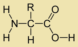
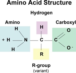
Amino-acids have a general structure that consists of an amino group, alpha carbon, carboxyl group and R’ chain( hydrocarbon attachment).

amino

The amino-acids exist as a charged species in ionic form or neutral species ( non-ionic). This depends on ?????, the charges present on the terminal ends of course. Compare these forms, the one above and the one below. Which is the ionic species?

amino

If you chose the first one then you won a prize of………..knowing the difference of a charge.

Wait there’s more…..

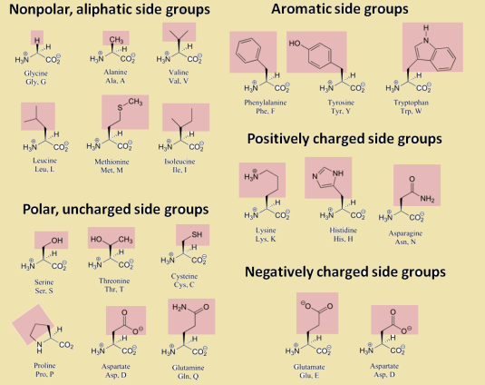
Amino acids have certain categories that exist in terms of its structure and their interacts with water and even themselves.

Amino-acids are unique in their structure but there exists various forms like polar uncharged R groups, non-polar  groups, positively charged, negatively charged and aromatic.

amino grps

Polarity and non-polarity are important in amino-acid structure and reactivity with each other.  Polar groups can interact with each other to form connective structures that leads the formation of hydrogen bonds with the amide group and carbonyl group.

More importantly amino-acids bond together to create peptide bonds or petide linkages to create a chain of amino-acids. That reaction is fueled by a condesation reaction. Note the positioning of the amino group and the carbonyl group.

meme6

Indeed Patrick, this helps in adding amino-acids to the peptide chain as more amino acids add to it. The carboxyl group must be exposed in order to react with the amino end of the adjacent amino-acid.

dipeptide-bond-formation

It’s a condensation reaction due to the fact water was lost from both structures combining. The important linkage is the bond of the carbonyl carbon of the carboxyl group to the nitrogen on the amino group. Hence the link is quite mutual in forming a polypetide chain.

Since on the topic of bond formation, other bonds do exist in the bonding of amino-acids. The disulphide bridge/linkage between two amino-acids call cysteine. The oxidation of two molecules allows for cystine to be formed and ” bridge” the gap. The reaction is reversible by reduction of the cystine to two individual cysteine molecules. This type of bonding is present in the structures of tertiary proteins in which we will see in oncoming posts.

cysteine

A dipetide is a chain formed between two amino acids and polypetide is between two or twenty amino acids long. Upon mentioning the number twenty there exists 20 esstential amino acids that you must obtain through dietary means.

meme8

To answer your question and that strange figure above amino acids can be obtained through everyday diet.  Meats and eggs are primary sources of amino-acids which are building blocks of proteins. For some of us that are a little reluctant of consuming meat,  beans and peas are alternative sources of amino acids. Plants which are our meatless……fat less…and minimal amino providing selection allow for creatine deficiency in our muscles. Creatine is an amino-acid that is a storage molecule for energy in our muscles and brains. Ever wonder about plants versus zombies, zombies eats plants to get to what?  the BRAINS of course. For that much needed creatine to keep those dead bodies moving.

pvz

meme9

Bear Grylls understands too much about proteins in his daring adventures, we know how he washes all of that down….. Since he resides in nature for all of us to experience the wild to our comfort with our cable television, air conditioning, lighting, bathrooms, internet and other household essentials he knows the natural proteins are the best.

Wait a hint has appeared remember for glucose that the D isomer is the most useful and exists commonly compared to the L isomer.

L isomers of amino acids are the common form compared to the D isomer. So the more natural the better. Won’t you say Bear….

your-father-is-full-of-proteins

Back to the Isomerism of the amino acids.

amide

The identification of the L isomer is determined by the placement of the atoms around the alpha carbon. The structures are arranged into the Fischer projections. The carbon molecules align themselves and the amino group assumes on the left or right of the chiral carbon. If the group is found on the left of the asymmetric carbon it is named L-isomer. So D isomer exists as the right…..

Functions Of Proteins.

They can acts as biological catalysts, storage molecules( ferritin), transport molecules of Oxygen and Carbon dioxide (hemoglobin), Protein pumps and protein channels and they assist in muscle growth. Due to different functional roles of proteins their structure differ from task to task.

ProteinStructure

There exist the primary(1) structure: that is in a linear arrangement and the amino-acids can be identified in a  particular order due the peptide bonds through out.

The secondary(2) structure consists of habitual folding patterns or an individual strain being warped around. They are the beta pleated sheet and the alpha helix. the beat pleated sheet can be represented by a pleated skirt or something like that and the alpha helix is like a telephone cord if got one that curls,  just so you know cordless phones have none.  The alpha helix contort to adjust themselves every 4th  amino acid to create a hydrogen bond of the electronegative oxygen on the carbonyl group with the hydrogen of amino group. These structures in alpha helices  can be disrupted by large hydrocarbon chains and other amino-acids.

phone card

The tertiary ( 3) structure of proteins are a little more complex with more bonding attributes like hydrogen bonding, salt bridges and disulphide bridges. This is essential for the tertiary  structures because various combinations allow for variety.

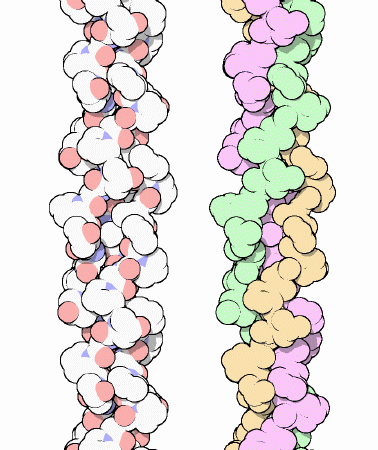
Structural proteins like Collagen:  Note the triple helix.

collagen

Example myoglobin for oxygen storage:

myoglobin

The Quaternary structure is a  much more complex arrangement. In these structures subunits can combine to form structures like haemoglobin with side chains and iron groups:

668px-Haemoglobin-3D-ribbons

As I come to a close I can share some interesting things I learnt in my lecture about Arginine, this amino-acid is responsible for the expansion of blood vessels. So it can assist for heart defects and help in blood pressure regulation. Gym buffs can utilize this for max reps.!!! .

meme5So certain amino-acids can be determined by the Ninhydrin test which produces a purple color.

aacid

Proteins can be determine by the biuret test that involves the reduction of copper(II) ions due to linking with the nitrogen. The basic solution of copper sulphate allows the hydrogen attached to the nitrogen to be removed.

In closing I leave this with you,

meme7References:

Elkaradagi S,Protein Structure and Structural Bioinformatics, http://www.proteinstructures.com/Structure/Structure/amino-acids.html(Feb,16,2014).

S. F. Betz,Disulfide bonds and the stability of globular proteins.,http://www.ncbi.nlm.nih.gov/pmc/articles/PMC2142256/(Feb,16,2014)

INTERESTING FINDINGSS MAN!

GLUCOSE OXIDASE SYSTEM  – SCIENCE BEHIND DIABETES TESTING

  • The measurement of blood glucose levels are achieved through the enzyme glucose oxidase.
  • The enzyme is only specific for D-glucose.
  •  Since oxidation can occur in glucose it is a reducing sugar
  • The enzyme glucose oxidase speeds up the oxidation of β- D- Glucose —> D-Gluconic acid. The α- D- glucose is quickly converted to the β form so that all the glucose can be measured one time.
  • Oxygen is the oxidizing agent acting upon the glucose. During the reaction the ring opens and the aldehyde group on C1 is converted into the carboxylic group (COOH) thus yielding the acid, D-Gluconic acid.
  • Simultaneously the oxygen and water react to form hydrogen peroxide. Another step is needed for the colour since the products are colourless.
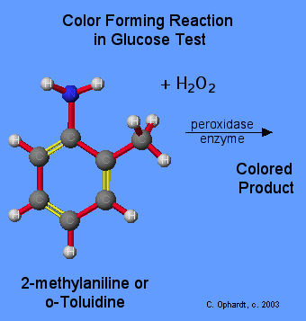
  • The H2O2 (hydrogen peroxide) reacts with a second colour producing chemical eg 2-methylaniline. It reacts with the H2O2 using the enzyme perioxidase to produce a coloured chemical.
  • The concentration of glucose is linked to the colour intensity. The more intense the colour, the higher is the glucose concentration.
  • BELOW SHOWS THE DIAGRAM OF THE GLUCOSE OXIDASE SYSTEM

548glucosetest 548glucosetest2

NITROCELLULOSE

The demonstration above deals with conversion, storage and release of energy. Using chemical means, we are changing ordinary cotton into explosive gun cotton. Cotton is made of cellulose. Please watch it the reaction is rather cool and exciting! You guys could probably try it out if you have the resources to your disposal! J

–          Concentrated sulphuric acid activates the nitric acid then nitrates the cellulose. Final product is trinitrocellulose which has –NO2 groups in place of the Oh groups

–          When the gun cotton touches a flame the following decomposition reaction takes place:

Nitrocellulose ——heat——–> CO2 (g) + CO(g) + H20(g) + N2(g)

–          The products of this reaction are all gases and therefore the reaction GOES POOF! WITHOUT ANY ASHES!tumblr_mhl7pqD07Z1qmjbn5o5_500

–          This is seen in magician’s  acts!

SADLY

We have now come to the end 😦

And you have learnt about carbs with enough knowledge to lend

If you liked this post and want to know more

Come back soon cause we’re gonna school you in biochemistry till you hit the floor! tumblr_n0fubz6Anr1rxivvco4_r1_250

See ya later biochemians! We are the twisted DNAs!

REFERENCES:

http://www.elmhurst.edu/~chm/vchembook/548bloodglucose.html

Click to access gun%20cotton.pdf

tumblr_makrvjHjVf1r9joxjo1_500

MR.CARBOHYDRATES PART 2

HEMIACETAL AND HEMIKETAL FORMATION

  •    Hemiacetal forms when an aldehyde becomes intoxicated with alcohol and the hemiketal is formed the same way except using a ketone.
  •     The two guys I’m going to use as examples are glucose and fructose.
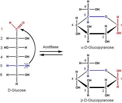
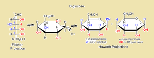
  •   Glucose cyclizes as its C1 aldehyde group reacts with a distal C5 OH to form a… you got it a hemiacetal! Intramolecular in fact, to form a 6 membered PYRANOSE ring.
  •  Fructose does the exact same thing except the C1 keto group reacts with C5 OH to form a HEMIKETAL! And a five membered FURANOSE ring is formedtumblr_mw5v5pN2hz1rthy8wo1_250
  •  Now now your pretty little heads are probably spinning right now! And you’re probably saying how is it that both are 6C sugars but one is 6 membered and one is 5? The thing is you name the ring not based on the # of carbons but on the numbers of sides.

These representations of the cyclic sugars are the HAWORTH PROJECTIONS. In which all the groups to the right of linear form is pointing down and all the groups to the left are pointing up in the cyclic projection.

glucopyranose

ANOMERS

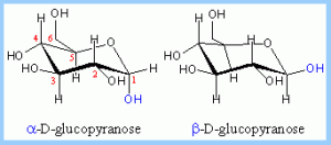
Because of cyclization of glucose it produces a new asymmetric center at C1. And the anomers α and β pop out because of this. Haworth projection result in planar rings with the OH either above or below the anomer C1. But how do tell the difference?

–          α– AB ALPHA IS BELOW

–          β – BA BETA IS ABOVE

n

SO HOW DOES THE STRUCTURE OF D-GLUCOPYRANOSE REALLY  LOOK?

“CHAIR” OR “BOAT” CONFIGURATION

Why do they call it chair or boat? Hmmm…let’s think maybe it looks like …….

boat-coloring-pages-16

images (3)

haworth

However we need to know which one is the stable configuration.  tumblr_inline_n0i5egQuTZ1rdaxds

To know which one will exist in nature duhh!!!

Kk let’s see the boat has two bulky groups pointing upwards along the same plane, wouldn’t these groups compete for space. Thus increasing steric hindrance and making the structure unstable.

download

However the chair conformations the carbons 2,3,5,6 lies in the same plane, C1 lies above and C4 lies below. The carbon atoms are antisocial they keep apart from each other at angles (110.90) thus increasing the space and reducing steric hindrance (electron repulsion) from the groups.

chair conformation of D-Glucose

chair conformation of D-Glucose

Hold up! There’s more..

In the chair conformation to determine whether either or is α or β LOOK AT THE OH GROUP!

–          OH GROUP POINTS DOWN- AXIAL POSITION – α

–          OH GROUP POINTS UP – EQUATORIAL POSITION- β

anomer

Ok Here we go again! Which one is more stable out of these two? tumblr_n0mh4lo7ko1sc1enmo1_500

The most stable conformation is the β chair conformation since it reduces steric hindrance or electron repulsion among the bulky groups.

MUTAROTATION

– Is the special rotation of a chiral compound due to epimerization. This is where the cyclic D-glucose when dissolved in water is rotate into their linear α and β forms and then the  β form rises and is seen in nature. The proportion of alpha to beta in equilibrium is 36% α and 64% β. WHY YOU ASK? Because the β  form is more stable since it reduces steric hindrance due to it being in the equatorial position or “up” position.

mutarotation1 mutarotation2 (1)

OH MR CARBS!!!

  • Two or more monosaccharides are joined by condensation reactions (it results in the loss of H20).
  • They are joined by a glycosidic bond- which is when C1 loses a H and let’s say C4 loses a OH to result in a loss of water. The C1 and C4 are joined by O. this is known as a (1-4) glycosidic bond.
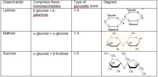
  • The following table shows the bonds between each three disaccharides:
  • disacc

Polysaccharides are formed in the same manner as disaccharides except with chains of monosaccharides. The table below summarises the three four main polysaccharides and compares the bonds between them.

–          A LITTLE RECAP THOUGH: starch is found in _____, glycogen in ______; cellulose in _____ cell walls. if you got it yourtumblr_ll880g1Q071qgkdixo1_500 a boss!

–          As seen in the table below glycogen has more branching than amylopectin (every 10 glucosyl residues).

–          This is because it permits rapid glucose release from glycogen stores eg in muscle during exercise. Animals are more active so they require a faster breakdown of glucose to gain energy.

–          Cellulose however has a zig- zag glycosidic bond in order to show that the second glucose molecule is flipped to facilitate bonding to C1 from C4.

table

Last but certainly not least………

REDUCING VS NON REDUCING SUGARS

–          A good question to ask is where is the reducing and non reducing end in a polysaccharide?

–          Well that’s easy the anomeric carbon that is not involved in the glycosidic bond is the reducing end  i.e the free anomeric carbon. However this only occurs where there is less branching.

–          You see the more branching you have——> the less anomeric carbons ——> the more non reducing you are.

–          C4- NON REDUCING END        C1- REDUCING END

–          Which one do you think will break down faster? Reducing or non reducing?

–          If you guessed reducing you areeeee RIGHTFULLLY WRONGGGG! THE ANSWER IS NON REDUCING. tumblr_n0ek8lNnpD1tn9hs9o1_500

Just think about it. If you have more branches wouldn’t enzymes want to bind to you to break you down faster than the reducing ends.

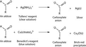
–          TEST FOR REDUCING AND NON REDUCING SUGARS

–          The Benedict’s test or Tollen’s (silver mirror) test. Below shows the equation for the reaction between benedict’s or tollen’s reagent with an aldose.

images (4)

Ok we are almost reaching an end I just have a few interesting facts for you. STAY TUNED GUYS! tumblr_inline_n0480tMxfi1s722jc

REFERENCES:

Click to access tutorial15.pdf

http://www.chem.ucalgary.ca/courses/351/Carey5th/Ch25/ch25-3-3.html

http://www.pslc.ws/macrog/kidsmac/toon_glu.htm

CARBS……CARBSS…..CARBOHYDRATES- the beginning

So essentially what fuels all the metabolic processes in our body? How do we go about our daily activities without passing out from the slightest movement? Yes, you’ve got it from the title, IT’S CARBOHYDRATES!

There is a huge knowledge base for carbohydrates and I will try to simplify most of the concepts and give you additional info to impress your classmates and lecturers. LET’S BEGIN!

carbs

Carbohydrates are macromolecules made of hmmm……

1) carbo- carbon

2) hydr-hydrogen, and well I’ll add it in

3) oxygen.

They are hydrates of carbon. Each are in equal proportions to form water. Their general formula is Cx(H20)y.

images

Carbohydrates play important functions or roles in living organisms such as:

1)      Energy source

2)      Storage

3)      Structure

4)      Precursor molecules

1)     ENERGY SOURCE

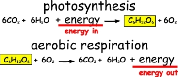
photosynthesis-respiration

  •  Just a small recap, since you all should know what both processes are: In photosynthesis Animated_bunny_hopping_aroundplants use CO2 and H2O to make glucose and oxygen. During respiration, animals break down glucose to CO2 and H2O.
  • Quick energy sources are the mono and disaccharides; these break down quickly. However the polysaccharides, starches take longer to metabolise.
  • The only polysaccharide humans cannot break down is…. CELLULOSE. This is because we lack the appropriate enzyme to break down the β(1-4) linkage.

2)      STORAGE

  • The main storage polysaccharides are starches and glycogen. Can you guess which is used by plants and animals?
  • Plants’ storage polysaccharide is STARCH while animals’ use GLYCOGEN.
  • The following chart below shows how carbohydrates in the diet is stored and glucose is oxidized to CO2 and H2O and energy (ATP) is released. The excess glucose is stored as firstly glucagon, then fats and amino acids.

Carbohydrate-function

3)      STRUCTURE

  • Cellulose plays a structural role in plants’ cell walls. It is a polymer of β-D- glucose and forms rigid and firm structure due to the fibrils, which makes it an excellent building material in plants. Its high tensile strength which allows it to stretch to withstand high hydrostatic pressures. In other words it stretches when the plant cells fills with water, so it would not burst.
  • Chitin is the structural polysaccharide of some insects’ exoskeleton. It is made up of β(1-4) linkages of the amino sugar N-acetyl-glucosamine which makes it very strong and flexible. It is used in medicine for sutures and is a strengthening agent for paper.

4)      PRECURSOR MOLECULES

  • Carbohydrates are precursors for the synthesis of certain biomolecules
  • Nucleic acids (De-oxy ribo nucleic acid and RNA) are composed of the carbohydrate ribose.
  • It also serves as raw materials for amino acid and fatty acid synthesis.
  • Disaccharides provide the building material for structures that protect the cell or whole organism.

Still don’t think carbohydrates are important. Imagine a world without carbohydrates; now imagine these aspects of life missing tumblr_n0hh9amAhJ1si5enjo1_500

–          No DNA or RNA

–          No bacterial cell walls

–          No plant cell walls and thus no plants

–          No energy stores, fuels or metabolic negotiators. Little to no metabolic reactions in the body due to no proteins and fats.

beast3

For my course I am only required to focus on the first process (THANK GOD!) however I will outline all for you. Carbohydrate metabolism involves the following processes:

1)      Glycolysis- Glucose-6- Phosphate —-à Pyruvic Acid

2)      Pentose phosphate pathway

3)      Glycogen synthesis and catabolism- glycogenolysis

4)      Gluconeogenesis- Lactic Acid—-à Pyruvic Acid —–à Glucose-6-Phosphate

The below diagram explains it perfectly…

590metabolism

SAC-A- WHAT ? WHAT? LOL… SACCHARIDES

  • Are sugars or starches
  • Consists of 2 basic compounds: aldehydes ( C=O) or ketones (RCOR)
  • There are various types however we will only focus on three main types:

1)      Monosaccharides– smallest sugar unit eg. glucose

2)      Disaccharides– two monosaccharides are covalently linked eg. sucrose

3)      Polysaccharides– a chain of two or more monosaccharides eg. starch

The diagram is a summary of each saccharide:

carbohydrate

As seen from the diagram monosaccharides are divided into two types based on the functional group:

1)      Aldoses(Aldehyde CHO

D-glucose_color_coded

D-Glucose

2) Ketoses (Ketone RCOR)

D-fructose

D-fructose

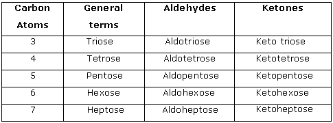
Monosaccharide are named judged solely by their number of carbons and whether they are an aldose or ketose! It’s that easy! The table below shows that:

monosaccharides-classification

SUGAR NOMENCLATURE scratch-head01-idea-animated-animation-smiley-emoticon-000414-large

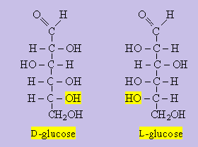
D VS L DESIGNATION ( Using Fishcer Projections) 

LOH on the  left  (L is for LEFT)

DOH on the right  ( D- don’t be a dummy, its RIGHT)

This applies to only chiral or asymmetric carbons( four different groups attached). To those sugars with more than one chiral carbon the D or L isomer is determined by the farthest carbon form the aldehyde or keto group. Still don’t understand watch the example below which are the FISCHER PROJECTIONS of D and L Glucose (linear forms).

epimers

EEEEKKK EPIMERS!!!

tumblr_ljxzu4dlPq1qgkdixo1_400

You probably wondering what ancient language I’m speaking, but not to worry I will explain epimerisation for you.

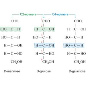
Epimers are two sugars in which their configuration differs around one carbon atom. Examples include D-Mannose and D- Glucose the OH differs at carbon 2 whereas with D- Galactose the OH differs at carbon 4.

The image below shows the differences between each Fischer projection:

epimers

Stay tuned for more we will be learning about the structure of these carbohydrates!… there’s still so much to learn! 🙂

REFERENCES:

http://www.tutorvista.com/content/biology/biology-iii/cellular-micromolecules/monosaccharides-and-oligosaccharides.php

http://www.medicalnewstoday.com/articles/161547.php


ALL ABOUT CELLS!

all about cells

Biochem has started off with a bang this year with the first topic covered being Cells! I thought I knew everything about cells, but the past week has proven me wrong! After attending my first two weeks of Biochemistry lectures I was thrilled to discover that there is so much more to cells than just the basic structure and functions. With a lecturer like mine (Mr. Jason Matthew), I’m sure that I know all there is to know about cells at this stage. I therefore recommend that you watch the interesting, informative video below to improve your knowledge of the cell.

cells

Fill in the blanks below by choosing the more suitable word from the bracket.          

There are two main types of cell.  ______________ (prokaryote/eukaryote) cells have a membrane bounded nucleus while _____________ (prokaryote/eukaryote) cells do not have nuclei. Prokaryotes have _______ (80s/70s) ribosomes and eukaryotes have _______ (80s/70s) ribosomes. Prokaryotes have ___________ (circular, simple/linear, complex) DNA while eukaryotes have __________ (circular, simple/linear, complex) DNA.

*NB*: ribosomes are measured in Svedsburg units, denoted by “s”, which is used to measure how fast molecules move within the cell.

c

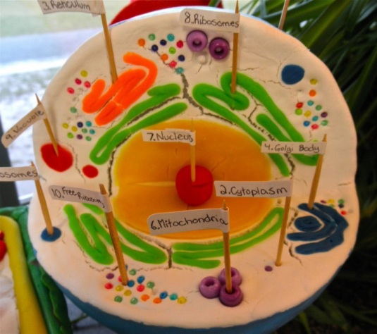
Figure 1- The cell model showing its structures. (msrosenreads.com)

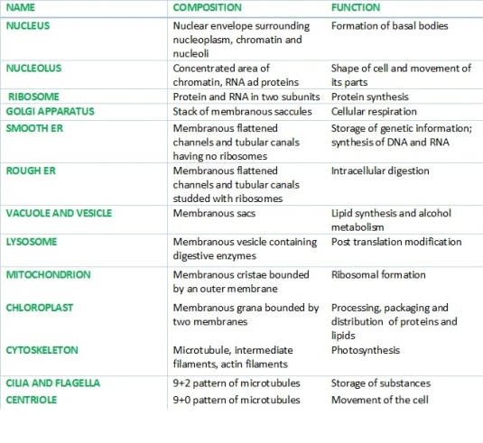
The table below shows the composition and functions of various structures of plant and animal cells. However, the FUNCTION column is not in order!! You must carefully read the contents of each column and match each structure to its corresponding function from the FUNCTION column. If you get all correct……you are a BIOCHEMATIC!

table

TABLE 1: SHOWING COMPOSITION AND FUNCTION OF VARIOUS CELL STRUCTURES

table

This is a simple concept by which the surface area of a cell is indirectly related to its volume. As cell size increases, the surface area to volume ratio decreases.

Many organisms have modified, both physiological and anatomical, to compensate for changes in the surface area to volume ratio associated with size differences.

EXAMPLES:

– The higher metabolic rates found in smaller (homeothermic) animals. Due to their large surface area relative to volume, small, homeothermic animals lose heat at much higher rates than large animals. Because of this, they are required to produce more heat to avoid the effects of thermal conductance.

– The variety of internal transport systems that have developed in plants and animals for actively moving materials throughout the organism, thus enabling them to circumvent the limits imposed by passive diffusion. Many organisms have developed structures that actually increase their surface area: the leaves on trees, the micro-villi on the lining of the small intestine, root hairs and capillaries, and the convoluted walls of arteries, to name a few.

table

I find this to be one of the most interesting topics while studying the cells!

Firstly, a cell containing multiple nuclei is called a SYNCYTIUM (plural- syncytia). Syncytia can be made by cell fusion as well as mitosis.

  • Most of our cells began with only one nucleus. However, some specific cells gain more nuclei upon maturing (e.g. skeletal muscle, osteoclasts), while others have no nucleus at all (e.g. red blood cells)!

Skeletal muscles begin as many single nuclei cells (myoblasts) and in maturity, fuse to form a single muscle cell or fibre that contains hundreds of nuclei from the original cells. These cells may need more nuclei because of their function. They are larger than other cells and constantly contract and relax by means of myofibrils which comprise of many different types of proteins, each with a specific task. This means that the skeletal muscle cells always require many proteins and we are aware that the nuclei are important in protein production since it contains DNA and transcription, the first step in making proteins, occurs in the nuclei.

o

  • On the other hand, each red blood cell forms in the bone marrow with a single nucleus that is expelled when the red blood cell matures in the marrow.

This leads to an interesting question: WHY ARE MATURE RED BLOOD CELLS WITHOUT NUCLEUS NOT PROKARYOTES?

The answer is simple to figure! Although red blood cells lack nuclei a maturity, they once had at the beginning of their formation, therefore they are eukaryotes. However, prokaryotes never have nucleus throughout their entire stages of development.

o

cel

Figure 3- simple diagram of cytoplasmic streaming. (bio1151.nicerweb.com)

  • Cytoplasm is made up of the cytosol as well as organelles, the cell’s internal sub-structures.
  • Cyclosis or Cytoplasmic streaming is the movement of the cytoplasm within a cell. Cytoplasmic streaming carries organelles and molecules (e.g. chloroplast, proteins and nutriens) from one part of the cell to another.
  • This circulation positions the chloroplasts so that they can absorb the maximum amount of sunlight for photosynthesis.
  • Cyclosis is a property of metabolically active, living systems. It does not occur in dead cells and microfilaments are responsible for the movement of Organelles and Vesicles in the Cytoplasm
  • The cytosol is the intra-cellular fluid of the cell containing different types of fibres called the cytoskeleton. which contributes to the cell’s shape, helps to move the organelles around within the cytoplasm.

cy

The cytoskeleton is a dynamic 3-dimensional structure that fills the cytoplasm, and is present in both eukaryotic and prokaryotic cells. The cytoskeleton acts as both muscle and skeleton, and aids in cell protection, cell motility (migration), cytokinesis, intracellular transport, cell division and the organization of the organelles within the cell.

In eukaryotic cells, the cytoskeleton is made up of the following three components:

1. Actin filaments (also called microfilaments): Monomers of the protein actin polymerize to form long, thin fibers that are about 8 nm in diameter. They provide mechanical strength to the cell, link transmembrane and cytoplasmic proteins, anchor centrosomes during mitosis, generate locomotion in cells and interact with myosin to provide the force of muscular contraction

2. Intermediate filaments: Cytoplasmic fibers about 10 nm in diameter. There are various types each constructed from one or more protein (e.g. keratins, nuclear lamins, neurofilaments, vimentins). All types of intermediate filaments provide a supporting framework within the cell

3. Microtubules: Straight, hollow cylinders about 25 nm in diameter, made of α-tubulin and β-tubulin dimers. They participate in a wide variety of cellular activities with most involving motion. Motion is provided by protein ‘motors’ that use the energy of ATP hydrolysis to move along the microtubule.

cy

cy

This topic was covered in the tutorial session and the information was well received by the entire class.

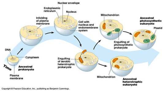
The endosymbiosis theory states that both key organelles (chloroplast and mitochondria) originated as free-living prokaryotic organisms.

 mito

Figure 4 – the origin of mitochondria according to the endosymbiosis theory. (http://www.origin-of-mitochondria.net/wp-content/uploads/28-04-eukaryoteorigin-l.jpg)

From the diagram above, the evidences of the endosymbiosis theory can be noted. They are as follows:

1.       The inner membrane is similar to the prokaryotic membrane while the outer membrane is similar to the eukaryotic membrane.
2.       It contains a circular DNA that matches up to that of prokaryotes. Eukaryotes have linear chromosomal DNA, not circular.
3.       Mitochondria divide by binary fission which is the same way in which prokaryotes divide. Eukaryotic cells divide by mitosis and meiosis.
4.       Mitochondria contain 70s ribosomes and prokaryotes also contain 70s ribosomes while eukaryotic cells contain only 80s ribosomes.
5.       Mitochondria was once free-living because for a cell to be free-living, it has to conduct basic metabolic processes. The mitochondria were well adapted to do this.

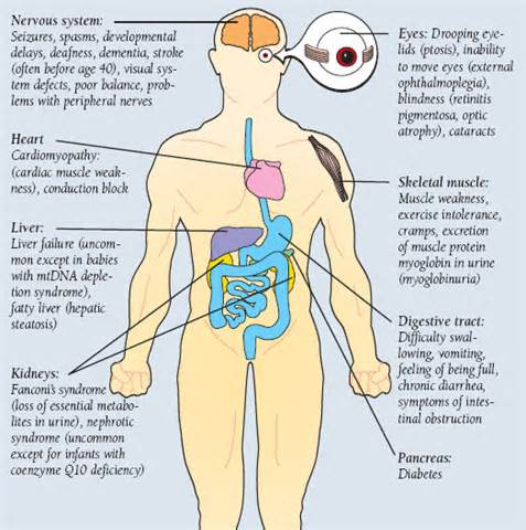
The mitochondrial DNA codes for some of the proteins needed for mitochondria production. Since the mitochondria have their own DNA mutations may result in mitochondrial diseases:

  • Mitochondrial diseases are the result of either inherited or spontaneous mutations in mtDNA or nDNA which lead to altered functions of the proteins or RNA molecules that normally reside in mitochondria.
  • Problems with mitochondrial function, however, may only affect certain tissues as a result of factors occurring during development and growth that cannot yet be explained.
  • Even when tissue-specific isoforms of mitochondrial proteins are considered, it is difficult to explain the variable patterns of affected organ systems in the mitochondrial disease syndromes seen clinically.

Different mutations in mtDNA and nDNA can lead to the same diseases.  In genetics, these are known as phenocopies.  A good example is Leigh syndrome, which can be caused by about a dozen different gene defects.  Leigh syndrome, originally a neuropathological description of the brain of one affected child, was described by physician Denis Leigh in 1951.

CHARACTERISTIC OF LEIGH SYNDROME:

  • Bilaterally symmetrical MRI abnormalities in the brain stem, cerebellum, and basal ganglia
  • Often accompanied by elevated lactic acid levels in the blood or cerebrospinal fluid.

CAUSES OF LEIGH SYNDROME:

  • NARP mutation,
  • MERRF mutation,
  • Complex I deficiency,
  • Cytochrome oxidase (COX) deficiency,
  • Pyruvate dehydrogenase (PDH) deficiency,
  • Other unmapped DNA changes.
  • Not all children with these DNA abnormalities will go on to develop Leigh syndrome, however.

In adults, mitochondrial diseases are more complex because detectable changes in mtDNA occur as we age and, conversely, the aging process itself may result from deteriorating mitochondrial function.  There is a broad spectrum of metabolic, inherited and acquired disorders in adults in which abnormal mitochondrial function has been postulated or demonstrated.

cy

body

Figure 5- types of mitochondrial diseases that affect the human body. (www.allmedicaldiseases.com)

Thank you for viewing the first of many interesting Biochem reflections. Twisted DNAs hope that it was of great help to you!

REFERENCES:

Video:

Text: