So long, farewell….

BrandnB: Okay guys, farewell till next time meaning a month or two till our next post… Ok you caught us… We will be back soon to share our understanding in the subject of Biochemistry. BrandnB here yet again expressing my thanks for the new followers, viewers, especially the viewers from other countries and also my fellow bloggers you know yourselves once again thanks till next time.

Hazel-Ann: Well guys this brings us to the end of blogging for this semester (don’t worry we’ll be back :P)! I must say the journey has just begun and so far it has been quite prosperous and I am honoured to have worked alongside all of the other members of Twisteddnas: Breaking the Bond! I have learned from this blogging activity that great team work is an important asset to a holistic individual. Constant blogging has also helped me in remembering what was taught by my amazing lecturer, Mr. Jason Matthew. Through this activity I was able to express my thoughts about Biochemistry thus far. I have also seen a great improvement in my creativity skills and have become more of an innovative thinker because of this blogging. I have learnt skills I have never been exposed to before like how to do a wordle, a video review, a reflection, even how to blog and much more! I am grateful for the experience and surely look forward to moving on to year two of Biochemistry and to embrace what it has to offer. I hope that Twisteddnas: Breaking the Bond has been helpful to you in many ways. So goodbye for now and I wish all of my colleagues good luck in final exams!

Kevin: Hello friends. This is Kevin here. It’s been a long and hard semester and we have finally completed our creative and helpful blog. This is definitely not a goodbye note but it’s simply a conclusion of all our work during this semester. I had absolute fun with this blog because it was a new, creative form of learning, not for just me but also for all our viewers and the other member of Twisted DNAs. I’d also like to thank our lecturer Mr. Jason Matthew for his guidance and effective teaching abilities. Additionally, I know I speak for everyone when I say that we all enjoyed making this blog and we hope you guys enjoy it as well. See you guys!!

Aaliyah: Hey guys! Well this semester has finally come to an end and that means blogging is over. *sad face* It definitely was an enjoyable experience, hard at times, but fun and helpful anyway. Continue to be the amazing Biochemians you are and work hard towards your goal! Thank you all for viewing our blog and I hope that it helped in some way J Farewell for now, wishing my peers (and myself lol) all the best in finals!

Kim: Hey guys its Kim. It has been great over the past few months blogging about biochemistry. I have learnt a lot while teaching the world biochemistry. Hopefully this is not the last time you guys hear from us. Certainly I have enjoyed blogging and so much so I have decided to take it up as a hobby of mine. Thanks guys for reading our blog and wish us luck for finals, they are in a couple weeks!


Youtube Review 2 – Glycolysis!

The video of review is a You Tube video entitled, “Glycolysis: The Reactions,” published by the NDSU VCell Production’s Animation. This five minute video covered the basic aspects of glycolysis such as what it does, where it occurs, the products of the reaction and the division into two phases. The video became more detailed as they included the ten reactions of glycolysis. For each reaction, they even named the enzyme catalyst, the products of the reactions and stated which reactions were irreversible. Additionally, chemical structures were provided in each reaction for better understanding of the changes that occur.
However, despite its great detail, the video failed to describe the importance of the some of the enzymes for example PFK-1 being of great significance with respect to regulation and Triose Phosphate Isomerase being a kinectically perfect enzyme. The video also lacked all the fates that the pyruvate molecule can undergo and the products of these fates. The only fate that was mentioned in the video was under aerobic conditions but no details were provided on this process. However, for a five minute video, it is quite detailed and helpful to persons trying to acquire a quick understanding of glycolysis. It is a video that I would highly recommend because of its organization and effectiveness in five minutes of teaching.
Kevin and Aaliyah signing out!

Video

MULTIPLE CHOICE QUESTIONS

For the following question ON ENZYMES:

Select the correct multiple answer using ONE of the keys A, B, C, D or E as follows:

A) 1, 2 and 3 are correct

B) 1 and 3 are correct

C) 2 and 4 are correct

D) Only 4 is correct

E) All are correct


A)    Which one is not a characteristic of competitive inhibition?

1)      The inhibitor binds to the enzyme-substrate complex

2)      It can be overcome by increasing the substrate concentration

3)      Km increases while Vmax decreases

4)      The inhibitor is similar in shape to the substrate


Multiple Choice 2 –Glycolysis

1)      For every glucose molecule entering glycolysis

a)      2ATP & 2NAD+ are used, 8ATP & 4NADH generated

b)      4ATP & 3NAD+ are used, 8ATP & 3NADH generated

c)      2ATP & 2NAD+ are used, 2ATP & 6NADH generated

d)     2ATP & 6NAD+ are used, 4ATP & 2NADH generated


YOUTUBE REVIEW 1

 Review of the video: LET’S TEST YOUR ATTENTIVENESS!!!

Hey guys this is Hazel-Ann and Kimberly here to bring you the YouTube review. So this video is sooo informative it’s just amazing…Mr. Jason Matthew basically explains everything you need to know about cells (at this introductory level of Biochemistry) in the most interesting, student friendly way possible. Apart from that, what I really love about this video is the presentation of the information. Mr. Jason ensures that he leaves you with visual representations of every topic he speaks about…so either way, it will be quite easier for the listener to recall what he has heard and observed. Below is a breakdown of information gathered from the entire video. We tried my best to present it in a creative, fun way to see how well both you and I have retained what we saw and heard in the video. The main points are listed below. I hope you enjoy it 🙂

-There are two main types of cell, prokaryotes and eukaryotes:
-Eukaryotes cells have a membrane bounded nucleus
-However prokaryote cells lack a nuclei.
-Prokaryotes have 70s ribosomes and eukaryotes have 80s ribosomes.
-Prokaryotes have circular DNA
-Eukaryotes have DNA in the form of linear chromosomes
-*NB*: ribosomes are measured in Svedsburg units, denoted by “s”, which is used to measure how fast molecules move within the cell.

Therefore we can officially say:

tumblr_n0m7dj0l0p1rgl5e0o1_250


Figure 1- The cell model showing its structures. (msrosenreads.com)

BLOG8

The table below shows the composition and functions of various structures of plant and animal cells. However, the FUNCTION column is not in order!! You must carefully read the contents of each column and match each structure to its corresponding function from the FUNCTION column. If you get all correct……you are a BIOCHEMATIC!

BLOG9


Also the differences between prokaryotes and eukaryotes were clearly stated in which the main distinction is whether the cell lacks a nucleus. The video was very informative and interactive which made me want to learn more. So I researched the cell and found out that mitochondria is inherited from the maternal line only! HOW COOL! Believe it or not we have our mother’s mitochondria. Mr. Matthews allows learning to be seen in a new light and actually makes you want to study. He explains everything thoroughly and allows you to answer questions to make sure you were not sleeping during the video or wake you up if you were. Lol just kidding.

Anyways guys that’s all about this youtube review! Hope you enjoy the video as much as we did!

bye2


PUBLISHED PAPER REVIEW 2.

Hello there!! This is Hazel-Ann and today I have a review of a published paper “Amino Acid Fingerprints Revealed in New Study” by Arizona State University. So it was a bright and sunny day and I decided to randomly browse the internet when BAM!!! I came upon the most interesting piece of science I have ever read! What I saw in this article augmented my knowledge in unexplainable ways. Yesss…I’m dead serious! So of course I couldn’t wait to share this new finding with you all.    

By now, the human genome project is no stranger to society. The human genome comprises three billion base pairs. Through the project, this code was decrypted effectively by 2003. This breakthrough in science has provided limitless insights and ideas pertaining to disease and the human health. If researchers can unlock the amino acid sequences from which protein is composed, scientists can have a better insight of diseases and the DNA. A group of researchers of Arizona State University led by Stuart Lindsay have embarked on an experiment to identify amino acids. Using state-of-the-art equipment, the team placed each amino acid between two electrodes and measured the current that went through each amino acid. Depending on this chain of current, which is characteristic of the amino acid, the scientists programmed a computer to identify each burst of electricity which indicated binding of an amino acid between the electrodes. Noise signals represented fingerprints and identified amino acids as well as modified variants. Currently, information on cancer, diabetes, Alzheimer’s and many other diseases are obtained from proteins. Protein sequencing will not only aid in patient treatment, but it will also be the means of advancement of molecularly examining the reaction of disease to therapeutics. Proteome is a huge amount of proteins.  Proteins are inevitably vital for growth, repair defense and catalysis. From the Human Genome Project, it was found that only 1.5% of genome codes for proteins. It is quite alarming that the human contains such a low gene number. But this can be accounted for by the modification of proteins generated from the DNA blueprint. It has been found that these proteins can alter their functions or become inoperable and this is detrimental to human health. This modification of proteins is caused by either alternative splicing (when exons are spliced and introns are cut off before translating into proteins) or post-translational modification (when markers like methylation and phosphorylation are added after production of proteins). Numerous cancers are linked with these modified protein faults. This makes for useful diagnostic indications. Once these proteins are identified, there will be a great revolution in Biomedicine. This led scientists to use the recognition tunneling method (used to determine the presence of particular nucleotide bases) for identifying the twenty amino acids in proteins instead of the four bases in DNA. This method has identified proteins that have been modified post transitionally from their unmodified precursor. As well as to distinguish between enantiomers and isobaric molecules (mirror image molecules). Lindsay states that many more researches and experiments are being executed for rapid progression of creating an amino acid fingerprints. This idea has solely been derived from the accomplishment of the human genome project. The hi-tech equipment are available for making this leap in Biochemistry and Biomedicine a successful one. Once the amino acid fingerprints are achieved, they can be applied to diagnostic medicine and can lead to a new era of research and findings for the cures of various diseases. The potentials of this project is limitless! So that’s it folks! Interesting, huh?? Can you imagine how easy it will be to diagnose diseases by knowing the amino acid sequence? What about using the fingerprints to find cures for various cancers? A new era of science dawns…


Reference:


Arizona State University. 2014. “Amino acid fingerprints revealed in new study.”                                                           ScienceDaily.www.sciencedaily.com/releases/2014/04/140406162416.htm (accessed April 8, 2014).

NUCLEOTIDES AND NUCLEIC ACIDS

Hey guys, Kimberly and Aaliyah here this week and we’re talking nucleotides and nucleic acids!

So we’ve learned before about the “central dogma” of molecular biology that basically encompasses the transcription from DNA to RNA and then the translation from RNA to protein.

Did you ever stop to think about what these things might be made of, however? Well today you’re gonna get the long-awaited answer for your question 😛

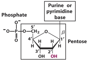
DNA and RNA are made up of nucleotides. These are the basic units or the building blocks of nucleic acids. Nucleotides are made up of three components- a sugar, a base and a phosphate group.

Components of Nucleotides:

Sugar: The first component we will talk about is the sugar part of a nucleotide. This is a 5-carbon sugar otherwise known as a pentose. Two kinds of these sugars are found in nucleotides. These are deoxyribose, which has a single hydrogen atom (H) attached to its carbon 2, and ribose, which has a hydroxyl group (-OH) attached to the carbon 2 instead. Deoxyribose is found in DNA while ribose is found in RNA. Deoxyribose gets its name simply because it is ribose that has lost an oxygen atom.


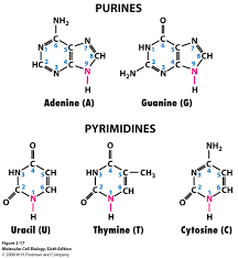
Base: The bases used in making up nucleotides are heterocyclic bases or nitrogenous bases (because they have a nitrogen incorporated into the ring, duh!). These bases can be separated into two types- the pyramidines and the purines. These have one ring, and are cytosine (C), thymine (T) and uracil (U), and two rings, and are adenine (A) and guanine (G) respectively. The pyramidines in DNA are cytosine and thymine and the pyramidines in RNA are cytosine and uracil, while the two purines, adanine and guanine are found in both DNA and RNA.

Phosphate: For nucleotide polymerization to occur and for these nucleic acids to be formed, a phosphate group must be present.

Altogether, these groups make up one nucleotide illustrated in the picture below.

When a pentose sugar is bonded to the base alone, the structure is called a nucleoside. When  phosphate group is added to the nucleoside, it becomes a nucletotide 🙂

DNAis made up of two polynucleotide chains, which are chains that are made by the joining together of nucleotide monomers that coil around each other. They not only coil around each other, but have bonds between the chains that hold the two polynucleotide chains together. This double helix is held together by hydrogen bonds that are formed between the bases of both chains. This occurs via complementary base pairing where adenine forms two hydrogen bonds with thymine and cytosine forms three hydrogen bonds with guanine.

It’s pretty simple once you see it in a picture isn’t it? Kinda looks like a twisted ladder, right? Lol Just think of the ladder rungs being the amine bases all paired up joined by hydrogen bonding, and the backbones being the sugar and phosphate parts! Easy like a Sunday morning.

Now where do bases attach to nucleic acids?

  • C1 position of ribose or deoxyribose
  • Pyrimidines attach via their N1 position of the ring to the pentose
  • Purines attach through the N9 position
  • Other bases(minor) have different attachments.
  • blog

ROLES OF NUCLEOTIDES

  • Building material of nucleic acids (RNA,DNA)
  • Provide energy in cellular metabolism (energy currency- ATP)
  • Allosteric effectors
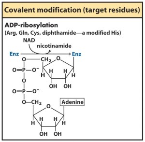
  • Enzyme cofactors are structurally made up of nucleotides eg NAD


ROLES OF NUCLEIC ACIDS

  • DNA contains:

1)      genes – information needed for functional protein and RNA synthesis

2)      promotors- segments which are involved in gene expression regulators

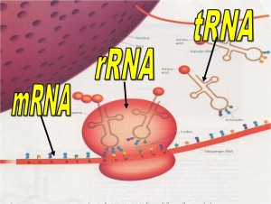
  • rRNAs (ribosome RNA) – made up of ribosomes which are involved in synthesis of proteins.
  • mRNAs (messenger RNA) – acts as a messenger owl carrying genetic information from gene to ribosome.
  • tRNA (transfer RNA) – information taken from mRNA is then translated into an amino acid sequence.
  • In some cases, RNAs perform catalysis.

Huh guys, imagine life without nucleotides or nucleic acids, do you think we would have even existed!  So I guess you can’t imagine life cause there would be none!


ATP

  • Adenosine triphosphate (nucleotide)
  • Contains adenine, ribose and triphosphate group
  • Energy carrier
  • Provides energy for other cells which perform jobs such as biosynthetic reactions, ion transport and cell movement.
  • Energy of ATP is made when the transfer of 1 or 2 of its phosphate groups to another molecule occurs. ADP + Pi —–à ATP
  • Reverse reaction is the hydrolysis of ATP to ADP.

http://hyperphysics.phy-astr.gsu.edu/hbase/biology/atp.html

NAD


NUCLEOTIDE REGULATION

  • COVALENT MODFICATION– activates some enzymes and inactivates others because enzymes are inactive until they are covalently modified so they could work. Eg Phosphorylation
  • blog2

NUCLEIC ACIDS

–          Polynucleotides

–          Oxygen at 5’ end of one nucleotide is connected to the oxygen of the 3’ end of another.

–         


NUCLEIC ACID STRUCTURE

–          The sugar phosphate is the backbone of the nucleotide

–          There are 2 separate strands:

1)      Parallel (3’à5’ direction)

2)      Antiparallel (5’à3’ direction)


–          Base pairing – hydrogen bonding holding the 2 strands together.

–         

–          http://www.visionlearning.com/en/library/Biology/2/DNA-II/160

This structure is important in:

–          DNA replication

–          RNA transcription

–          Backbones are the sugar –phosphate which are negatively charged so it is located outside


–          Planner bases are stacked above each other like pancakes however on the inside


HOW ARE NUCLEIC ACIDS FORMED?

–          Monomers of nucleotides are joined by phosphodiester linkage which is a formation between the hydroxyl (OH) group at the 3’ end of nucleotide and phosphate of a next nucleotide.

–          The 5’end then lacks a nucleotide at the 5’ position and the 3’ end lacks a nucleotide at the 3’ position.


HELICAL TURN

–          10 base pairs per turn

–          34 amino acids per turn

–          There are 3 helices called:

1)      A form

2)      B form

3)      Z form

blog3

NUCLEIC ACIDS

–          Most common form for DNA is the B form. It is the standard DNA double helix.

–          Most common form for RNA is the A form. WHY?

–          Because of its deeper minor groove and shallow major groove. It is also favoured in low water conditions.

–          Z form:

–          Has a narrow, deep minor groove

–          Has no major groove

–          Can form some DNA

–          However requires alternating syn and anti base configurations

–          It is a laboratory anomaly

–          Because of its high salt or charge neutralization and it is left handed while most helices are right handed.

B- DNA

–          Has a deeper major groove  and a

–          Shallow minor groove


RNA

–          rRNA – RNA + PROTEIN -à RIBOSOMES

–          mRNA

o   carries DNA code to cytosol for protein synthesis or ribosomes.

o   Are read in codons  or triplets from (5’ to 3’ direction)

–          tRNA

o    carries amino acids to mRNA and like a puzzle matches up by base-pairing of anti codon with mRNA codon.

blog4

STABILITY OF NUCLEIC ACIDS

There are many factors which stabilize and destabilize the helix of nucleic acids. They are:

1)      HYDROGEN BONDING

o   Contributes to DNA double helix, RNA secondary structure

2)      STACKING INTERACTION OR HYDROPHOBIC INTERACTION BETWEEN BASE PAIRS

o   It is energetically favourable

o   Maximized stacking in double-stranded DNA


EFFECT OF ACID ON NUCLEIC ACIDS

–          Strong acid and high temperatures

o   They are hydrolysed to base,riboses  or deoxyriboses and phosphate.

–          At pH 3-4 apurinic nucleic acids are formed. This is due to the glycosylic bonds attaching the purine A and G bases to the ribose ring are broken.

–          However formic acid regenerates this.


EFFECT OF ALKALI ON NUCLEIC ACIDS

–          HIGH pH (>7-8)

o   Small effects on DNA structure

–          HIGH pH

o   Changes the tautomeric (keto and enol forms) state of bases.

–          Change in tautomeric states of bases results in DNA denaturation therefore it results in unstable base pairing.

blog5

–          RNA  is unstable at higher pH because of its 2 OH groups.


CHEMICAL DENATURATION OF NUCLEIC ACIDS

Two chemical which denature nucleic acids are:

-Urea (H2NCONH2)

– Formamide (HCONH2)

HOW IT OCCURS?

-Disrupts hydrogen bonding of water solution

-Reduction of hydrophobic effect between bases

-Denaturation of strands in double helix


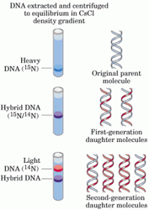
BUOYANT DENSITY OF DNA

–          1.7gcm-3 = 8M CsCl

–          It is used in DNA purification

blog6


C3 SPECTROSCOPIC AND THERMAL PROPERTIES OF NUCLEIC ACIDS

1)      UV SPECTROSCOPY

o   Absorption of UV light due to bases

o   Maximum wavelength absorption by both DNA and RNA (Max=260nm)

o   Applications: detection, quantitation , assessment of purity (A260/ A280)

2)      HYPOCHROMICITY

o   Creation of hydrophobic environment by stacking of bases making them less accessible to UV absorption (dsDNA, ssDNA or RNA nucleotide)

o

3)      QUANTITATION OF NUCLEIC ACIDS

o   Extinction coefficient- 1mg/ml dsDNA has an A260 of 20 ssDNA and  25 RNA

o   The values for ssDNA and RNA are approximate

o   Values are the sum of absorbance of different bases (purines are more than pyrimidines)

o   Absorbance values depend on the amount of secondary structures due to hypochromicity or stacking of bases.

4)      PURITY OF DNA

o   A260/ A280

o   DsDNA – 1.8

o   Pure RNA -2.0

o   Protein – 0.5

5)      THERMAL DENATURATION/MELTING

o   Destruction of the double stranded hydrogen bonded regions of DNA and RNA

o   RNA – absorbance increases gradually and irregularly

o   DNA- increases cooperatively.

o   Tm- melting temperature. This the temperature at which a 40% increase in absorbance is achieved

blog7


6)      RENATURATION

o   Rapid cooling- only allows formation of the local base pairs and absorbance is slightly lowered.

o   Slow cooling- the formation of the whole complementation of dsDNA and absorbance decreases gradually and cooperatively just like denaturation

OK guys i think that’s enough for nucleotides and nucleic acids. SEE YOU NEXT TIME! have a great weekend!

tumblr_n06sc0RY9Z1ramgxlo1_400


LIPIDS

7p0yb

 As the above picture indicates today’s topic gonna be discussed are lipids.

So BrandnB and Kevin are here to produce some knowledge about what we learnt in the past week about lipids.

Let’s Begin…..

Lipids are sources of energy, performs structural functions, cell signaling and identification, insulation  and to create cell membranes.

Lipids produce more energy per gram  because they are more reduced as compared to hydrocarbons. Fat has to be hydrolyzed into the fatty acid and alcohol with then takes alternate pathways to be utilized to create energy.

Lipids have a common name but are classified into several groups.

  • Fatty acidsfatty acids
  • Phospholipids

phospholipids

  • Triacylglycerol

trigyl

  • Steroids

steroids

  • Gylcolypids

gyl

Let’s get at it.

Lipids can be saturated and unsaturated, they posses carbon to carbon single bonds and carbon to carbon double bonds respectively.  These properties can lead to variation in  physical properties.

fat_vs_oil

As you can see the differences in saturation and unsaturation are the presence of the C=====C .

7p3g0

Saturation deals with the functional C—-C or C====C bonds. Therefore the physical properties vary such as state at room temperature, melting point, solubility in some cases.

Fats are saturated  and generally solid at room temperature  which allows then to  posses a higher Melting point than oils due to the lack of whereas oils are unsaturated and liquid at room temperature. Lets begin who will win.

butter-oil

As we know butters and margarine are bad for you as well as excess oils. There exist a problem of the lesser of two evils. Fats allow foods to taste extremely better due the ability carry fat soluble vitamins such as vitamin A,D,E and K. But it leads to Plaque build up in our circulatory system and Heart Diseases. The figure below shows a normal artery , artery with plaque build up and finally a clogged artery. This is due to the deposit of  lipids in the artery walls that beings to block the lumen the eventually leads to atherosclerosis and Coronary heart disease.

arteries-clear-and-clogged7p12r7p14u


Well I guess she found out that grandma use to put 3 sticks of butter in  her cakes to give that special taste, we know how granny makes  it.

Did you know Beeswax is created from lipids, by reacting a fatty acid and a long chain alcohol.

beeswax

Bees and Ants also use a mono-saturated fatty acid (oleic acid) to signal to remove dead bodies from an area.  This chemical signals other worker ants or bees to remove these bodies because they do not want to pollute the nest or infect their queen.

oliec acid


The structure above is cis-oleic acid.

Since We figure that you might think that Hey is this similar to amino acids with the carboxyl group on the right and in this case a methyl group on the left.

7p0z5This particular orientation allows for Designation.

But for assignment of the omega,  monosaturated and unsaturated fatty acids we start form the methyl end and the first point of unsaturation.

As we know not all fatty acids are made in the body but can be obtained through diet as anything else essential due to the body being unable to synthesis a C===C before the 9th Carbon.

Therefore Omega 3 and Omega 6 fatty acids must be obtained through diet.

omega3

Aside

TCA AND ETC

Hey guys, this is Hazel-Ann and Aaliyah and we’ll be taking over this week. We’re so excited to talk about the TCA and ETC! Week 8 has truly been interesting since we had only quizzes during our lecture time…YIKES!!!!!

Okay so back to the topic….TCA: have you ever wondered why it’s called TCA instead of TAC? I mean after all it stands for Tricarboxylic Acid Cycle right??

Baffled yet??…wait there’s more: this cycle actually has two other names: Citric Acid Cycle and Krebs Cycle!

The Citric Acid Cycle is a sequence of enzyme-catalyzed chemical reactions which are imperative in aerobic respiration in cells.

  • The cycle begins with the end product of glycolysis (the first step of cell respiration), pyruvate.
  • In glycolysis, glucose is decomposed into pyruvate.
  • A two carbon fragment of pyruvate is used to form acetyl coenzyme A. The acetyl-CoA enters the Krebs cycle which occurs in the mitochondria.
  • In the process of converting pyruvate to acetyl-CoA, carbon dioxide is produced and a molecule of NADH is formed.

Step 1:

A molecule of citrate is formed by the joining of the Acetic acid subunit of acetyl CoA with oxaloacetate.  The function of acetyl coenzyme A is to transport acetic acid from one enzyme to another. After this, the Krebs Cycle is restarted when hydrolysis releases the coenzyme so it may combine with another acetic acid molecule.

Step 2:

Then, citric acid molecule goes through an isomerization.  Water is removed from the citrate structure but a double bond forms between the 2 carbons and the water molecule is reintroduced. However, unlike the first citrate molecule structure, the hydroxyl group and hydrogen molecule are transposed. Hence, Isocitrate is forms.

Step 3:

Oxidization of the isocitrate molecule occurs by a NAD molecule that is reduced by the H atom and the OH group. NAD binds with a hydrogen atom and carries off the other hydrogen atom leaving a carbonyl group.  Due to the instability of structure a CO2 molecule is released forming alpha-ketoglutarate.

Step 4:

Here, coenzyme A, reappears and oxidizes alpha-ketoglutarate.  NAD reduction occurs again producing NADH and another H atom remains. Again, a carbonyl group is released as carbon dioxide and is replaced by a thioester bond between the previous alpha-ketoglutarate and coenzyme A while a molecule of succinyl-coenzyme A complex is produced.

Step 5:

Hydrogen atoms from a water molecule are attached to coenzyme A. Coenzyme A is then displaced by a free phosphate group which transfers to a GDP molecule to yield a GTP energy molecule. A succinate molecule remains.

Step 6:

Flavin adenine dinucleotide (FAD) oxidizes succinate removing two H atoms from it and forms a double bond in between the two C atoms, making fumarate.

Step 7:

Malate is formed by the addition of water to the fumarate by an enzyme.

Step 8:

An NAD molecule oxidizes the malate molecule. The OH carrying carbon is transformed into a carbonyl group.  Oxaloacetate is the end product. It can then join acetyl-coenzyme A and resart the Krebs cycle.

Summary:

In summary, Krebs cycle involves 3 major activities.

1)      Production of 1 GTP (guanosine triphosphate) which eventually donates a phosphate group to ADP to form 1 ATP;

2)      Reduction of 3 NAD molecules;

3)      Reduction of 1 FAD molecule.

In the cell’s energy-generating process, formation of reduced NAD and FAD are more imperative than 1 molecule of GTP producing 1 ATP because NADH and FADH2 give their electrons to the electron transport chain (ETC) that creates a lot of energy by making a lot of molecules of ATP.

http://www.incolor.com/mcanaday/Krebs%20Phases.htm

If it’s too much to digest right now how about this?

I KNOW RIGHT! 

So let’s move on!

IT’S TIME TO HOP ON THE ELECTRON TRANSPORT TRAIN….NO….CHAIN

Now, the electron transport or respiratory chain is where electrons are transported to join oxygen from respiration at the end of the chain. The overall ETC reaction is:

2 H+ + 2 e+ + 1/2 O2 —> H2O + energy

Pre-Initiation of Electron Transport Chain:

Figure 3: simplified illustration of ETC

ETC is started by oxidation reaction of an organic metabolite with the coenzyme NAD+ (nicotinamide adenine dinucleotide). 2 hydrogen atoms are removed from the organic metabolite which is usually from the citric acid cycle and fatty acids oxidation:

MH2 + NAD+ —–> NADH + H+ + M: + energy, where M= any metabolite (usually an alcohol oxidized to a ketone).

Removal of one hydrogen with 2 electrons as a hydride ion (H) and another as the positive ion (H+) occurs.

NAD+ is a coenzyme containing the B-vitamin, nicotinamide.

The other steps in the ETC are:
1) to pass along 2H+ ions and 2e to eventually react with oxygen;
2) to save energy by forming 3 ATPs; and
3) to restore the coenzymes back to their original form as oxidizing agents.

Initiation of Electron Transport Chain:

  • NADH interacts with the first complex 1 enzyme, known as NADH reductase. Complex 1 contains a coenzyme flavin mononucleotide (FMN), similar to FAD.
  • The NADH, and another hydrogen ion then enter the enzyme complex and pass along the 2 hydrogen ions into the mitochondrial interspace. Acting as a pump, these H atoms are used by ATP synthetase to form an ATP for every two hydrogen ions produced.
  • Performing similar to this, three complexes (1, 3, and 4) produce 2 hydrogen ions each, therefore 3 ATP are produced for every use of the complete ETC.
  • NADH passes along 2 electrons to FMN, iron-sulfur protein (FeS), and lastly to coenzyme Q. These reactions restore coenzyme NAD+ and create a cycling effect. The NAD+ is then ready to react further with metabolites in the TCA cycle.
  • Coenzyme Q makes CoQH2 by joining 2 H ions and is soluble in the lipid membrane and moves through the membrane to encounter enzyme complex 3.
  • Enzyme complex 1 of the ETC is combined with the formation of ATP:

a) MH2 + NAD+ —> NADH + H+ + M + energy

b) ADP + P + energy —> ATP + H2O


Figure 4: Illustration of ETC in mitochondria and the association with TCA Cycle.


Enzyme Complex 3:

  • Cytochrome reductase bc is a series of activities through enzyme complex 3 that begins when CoQH2 carries 2 extra electrons and 2 extra hydrogen ions.
  • Cytochromes are similar in structure to myoglobin or hemoglobin. The heme structure containing the iron ions, initially in the +3 state and changed to the +2 state by the addition of an electron is significant. The CoQH2 passes along the 2 electrons first to cytochrome b1 heme to b2 heme, to an iron-sulfur protein, then to cytochrome c1, and finally to cytochrome c.
  • The 2 hydrogen ions are directed to the inside of the mitochondria for conversion into ATP.


Figure 5: Illustration of cytochrome b, c complex 3.

Complex 4:

  • Cytochrome c is a small molecule able to travel in the lipid membrane layer and diffuses toward cytochrome a complex 4.
  • The transport of the electrons continues, and this is the third and last time that 2 hydrogen ions are directed to the inside of the mitochondria for ATP conversion.
  • ATP synthetase is found throughout the bilayer membrane of the mitochondria. The pumping of the re-entry of the hydrogen ions through the ATP synthetase produces 3 ATP.
  • Finally, diffusion of oxygen into the cell and mitochondria occurs for the last metabolic reaction. A water molecule is produced by reaction of oxygen atom with the 2 electrons and 2 hydrogens.

http://www.elmhurst.edu/~chm/vchembook/596electransport.html

INTERESTING FACTS ABOUT TCA AND ETC

  • The tricarboxylic acid cycle got the common name “Krebs’ cycle” after the biochemist Hans Adolph Krebs when he discovered the steps of the cycle through a series of experiments, in 1937.
  • This cycle is the second of three parts in the process of breaking down glucose into energy that the body can use, in the form of ATP. The stage before TCA is glycolysis and the stage after is oxidative phosphorylation.
  • The Electron Transport Chain (ETC), while it is the last step of the three in gaining energy from the foods we eat, it is actually THE most important step since most of the ATP is produced here.
  • In glycolysis, the final net production of ATP molecules is 2, in the citric acid cycle, it is 2 and in the electron transport chain, the net production of ATP is 32 ATP molecules. In total, 36 ATP molecules are formed from the entire process during aerobic respiration per pyruvate molecule.
  • In ETC electrons move from high energy to low energy making a proton gradient that then pushes the production of ATP.
  • Altogether TCA and ETC are ESSENTIAL for human survival, since these stages to produce ATP give us energy to carry out our daily activities. ATP is also important in synthesizing RNA and DNA.

So that’s all for this week peeps…thanks for tuning in, hope that this was helpful! Don’t forget to check us for our next post, next week same time.

REFERENCES:

http://www.elmhurst.edu/~chm/vchembook/596electransport.html

http://www.incolor.com/mcanaday/Krebs%20Phases.htm

https://highered.mcgraw-hill.com/sites/0072507470/student_view0/chapter25/animation__how_the_krebs_cycle_works__quiz_1_.html

http://hyperphysics.phy-astr.gsu.edu/hbase/biology/tca.html

THE LINK BETWEEN OBESITY AND FRUCTOSE METABOLISM

Hey guys this Kim and Kevin here to bring to you part of glycolysis.

I essentially wanted to show you how biochemistry can tie in with real life problems that many people especially children suffer with. OBESITY!

Image

Image

Obesity contributes to all these factors, not just being obese.

<<<< SAY U LOVE ME LIKE A FAT KID LOVES CAKE! Ok that was a little humour but seriously this is an important issue that is threatening kids’ lives today!

So just to juggle your mind fructose is one of the monosaccharide polymer of the disaccharide sucrose ( table sugar). It is wayyyy sweeter, more soluble than sucrose and inexpensive to produce in the form of high fructose corn syrup. Well you know this just entices manufacturers of sodas and processed foods. However those of you chefs out there that like to sweeten with this and even those who consume these foods concentrated with it, there are many dangers of high fructose corn syrup which are listed below:

Image

 Alright let’s try to link it to obesity

Obesity is a very serious epidemic. One of the factors that may be contributing to this epidemic is consuming way too much high fructose foods and drinks. So guys you see all those Coca- cola drinks and canned syrups and fruits THOSE ARE A BIG NO-NO! This is essentially what is in 20 ounce glass of coca cola.

Image Image

Let me show you why through the explanation of the metabolism of fructose.

There are 2 ways fructose can be metabolised in the body: (which parts of the body do you think these occur?)

  • Adipose tissue, muscle and kidney- the enzyme hexokinase phosphorylates or converts fructose into fructose- 6-phosphate and then enters glycolysis. Below shows the what happens in equation form:

Fructose + ATP ——-à Mg2+  fructose-6-phosphate + ADP

  • Liver- this is the pathway that fructose is primarily metabolised since hexokinase is not present but the enzyme glucokinase. Glucokinase has a low affinity for fructose or basically hates and avoids fructose. Therefore fructose goes through the fructose-1-phospate pathway. Below shows the summary of fructose metabolism in the liver:
  • Image


Reasons for high fat storage:

1)      – Fructose is phosphorylated to give fructose- 1 – phosphate by enzyme fructokinase. It’s that easy just remember 1 goes in between fructose and phosphate.

–          Then the fructose- 1- phosphate is then split up by the enzyme fructose-1 – phosphate aldolase into two, 3C molecules ( glyceraldehyde and dihydroxyacetone phosphate) Gosh I know right those words are so long to remember so here’s an acronym for the 2nd one (DHAP)

–          Only one the 3C molecules actually move on to the 2nd stage of glycolysis. Which one do you think it is? Don’t worry if you got it wrong, this question is tricky! It’s DHAP! It can be converted by the enzyme triose phosphate isomerase to glyceraldehyde-3-phosphate and then it continues into glycolysis.

–          However the glyceraldehyde is converted into glyceraldehyde-3-phosphate which is then converted to glycerol- 3- phosphate. And why do you think this latter conversion is bad? Ok let’s try to break it down where have you seen the word glycerol already, not in triglycerides which are mainly lipids(fats). However in this case it contributes to triacylglycerols. This can cause more fat being stored thus more fat + no exercise———à OBESITY

2)      The second reason for the contribution of the obesity epidemic is that:

–          There is a step that fructose metabolism completely bypasses in glycolysis which is the phosphofructokinase-catalyzed step and thus it avoids a major regulatory point.

–          Therefore disruption of the metabolism occurs and there is a greater lipid production or more fat is produced than in glucose metabolism.

–          This is why eating too many high fructose foods result in obesity due to fat storage.

Below shows a summary of the metabolism of fructose as well as glucose which has been discussed before:

Image

Here is a simpler diagram to follow from:

Image

So next time you want to pick up that can of coke or partake of  a McFlurry or even Grandma’s sweet homemade cookies just think again before it leads to this:

Image

Alright see you next time guys, talking to you about this topic has been informative and fun for both you and I. Take care, see you next time!

Image

REFERENCES:

BOOKS:

Pratt, Charlotte W., and Kathleen Cornely. Essential biochemistry. Hoboken, NJ: J. Wiley, 2004.

WEBSITES:

https://www.ucsf.edu/news/2009/06/8187/obesity-and-metabolic-syndrome-driven-fructose-sugar-diet

http://biochemistrystudy.blogspot.com/2012/10/feeder-pathways-for-glycolysis.html

Glycolysis, Parts 1 and 2

shibe-meme-learning

Hey guys, Kimberly and Kevin are here with you this week and the topic of discussion is Glycolysis. For those of you who may consider this topic as equally horrendous as calculus, it truly isn’t because I too, was under that impression and you will soon realize the simplicity once you complete our fun and interactive blog on Glycolysis. Enjoy!!

Before we can truly learn about Glycolysis, we must understand the basics:

  • Glycolysis can be described as the oldest of all metabolic pathways.
  • It takes place in ALL life.
  • It occurs in the cytoplasm of the cell.
  • It forms 2 pyruvate molecules from the breakdown of a glucose molecule.
  • It is NOT oxygen dependant, that is, it can occur in an anaerobic environment.
  • It produces a net gain of 2 NADH molecules AND 2 ATP molecules per glucose molecule.

Image

Lysis means “to split.” Therefore, Glycolysis can be easily remembered as the splitting of glucose which is a 6 carbon structure into two pyruvates which are 3 carbon structures.

Image

The Glycolysis process above can be divided in TWO phases:

1.) The Energy Investment Stage (Preparatory Stage) in which 2 ATP forms 2 ADP and occurs from step 1 to 5.

2.) The Energy Generation Stage (Payoff Stage) in which 4 ADP forms 4 ATP, 2 NAD+ forms 2 NADH and occurs from step 6 to 10.

Image

If this is your reaction at the moment, then DO NOT PANIC! It gets easier after the following diagrams and explanations, so keep calm and LEARN GLYCOLYSIS! 😀

1.) Preparatory Stage – conversion of glucose to glyceraldehyde 3 – phosphate via phosphorylation.

  • Image

The numbers 1 to 5 represent the specific enzymes present as illustrated in the diagram above:

  • Enzyme 1 = Hexokinase; phosphorylates the glucose molecule to form glucose 6 – phosphate.
  • Enzyme 2 = Phosphohexose Isomerase; helps convert glucose 6 – phosphate to fructose 6 – phosphate.
  • Enzyme 3 = Phospho – fructokinase – 1; most important enzyme in terms of regulation.
  • Enzyme 4 = Aldolase; helps split the 6 carbon sugar phosphate to form the 3 carbon sugar phosphates.
  • Enzyme 5 = Triose Phosphate Isomerase; converts DHAP molecule into G3P molecule.

2.) Payoff Stage – conversion of G3P to pyruvate via oxidation and the formation of both ATP and NADH

Image

The numbers 6 to 10 represent the specific enzymes present as illustrated in the diagram above:

  • Enzyme 6 = Glyceraldehyde 3 – phosphate dehydrgenase; catalyzes the oxidation of the aldehyde group to a carboxylic group and phosphorylates the G3P to 1,3 – BPG
  • Enzyme 7 = Phosphogylcerate kinase; 1,3 BPG loses a phosphate group which is transferred to an ADP molecule to form ATP
  • Enzyme 8 = Phosphoglycerate mutase; phosphate group on carbon 3 is removed and the one on carbon 2 remains.
  • Enzyme 9 = Enolase; dehydrogenation occurs.
  • Enzyme 10 = Pyruvate kinase; form ATP when converting PEP to pyruvic acid.

Image

Have no fear my viewers because we’re almost at the end!! Let us move on to the Fates of Pyruvate. The following diagram is a simple illustration showing the different fates of pyruvate.

Image

The following table represents the same concept as the previous diagram, however the structural formulas of the various conversion reactions of pyruvate are illustrated in this table.

Image

1.) Aerobic Conditions – Entry into TCA cycle.

  • The pyruvate is converted to Acetyl – Co A via an oxidative decarboxylation reaction.
  • The enzyme that catalyses this reaction is pyruvate dehydrogenase.
  • This reaction can only occur if mitochondria are present.
  • This reaction is referred to as a link reaction because it links pyruvate to the TCA cycle.


2.) Anaerobic Conditions – Lactic acid Fermentation

  • The pyruvate is converted to lactate.
  • The enzyme that catalyses this reaction is lactate dehydrogenase.
  • Lactic acid fermentation regenerates NADwhich allows glycolysis to be completed as there are limited NAD+ molecules within a cell and is important for the erythrocyte.
  •  No ATP is generated when pyruvate is converted to lactate.


3.) Anaerobic Conditions – Alcoholic Fermentation

  • This is a 2 step reaction that first converts pyruvate to acetaldehyde and then to ethanol.
  •  The two enzymes that catalyse these reactions are pyruvate decarboxylase and alcohol dehydrgenase.
  • Pyruvate decarboxylase requires TPP as a cofactor for the enzyme to work.
  • Alcholic fermentation regenerates NADwhich allows glycolysis to be completed as there are limited NAD+ molecules within a cell.


AND THAT’S IT FOR MY PART GUYS!!!! Until next time.


g1332380942943969136

References:
Avril Chew. 2003. GCE ‘A’ Level Biology @ Your Fingertips. Singapore : Redspot Publishing. (accessed March 9th, 2014)

CJ Clegg with DG Mackean. 1994, 2003. Advanced Biology Principles and Applications. 2nd edition. London : Hudder Education. (accessed March,9th,2014)

BiochemJM You Tube Page. Glycolysis Parts 1 and 2.

http://www.youtube.com/watch?v=K-NMuq-XIHo&feature=youtu.be
http://www.youtube.com/watch?v=33JUjeo6-lE (accessed March 9th, 2014)

Previous Older Entries